Figure 1.
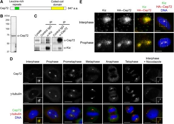
Cep72 associates and co-localizes with Kiz at and around the centrosome. (A) Schematic diagram of the Cep72 protein. Cep72 has two leucine-rich repeats (green, amino acids (a.a.) 55–76 and 77–98) and a potential coiled-coil domain (yellow, a.a. 476–620). (B) Western blotting of HeLa cell lysates for Cep72. (C) HeLa cell lysates were immunoprecipitated with control IgG or with anti-Cep72 or anti-Kiz antibodies. The co-precipitated proteins were analyzed by immunoblotting using the indicated antibodies. (D) Immunostaining of HeLa cells for Cep72, γ-tubulin, and DNA. (E) To examine the co-localization of Cep72 and Kiz, HeLa cells were transfected with an expression vector for HA–Cep72, fixed, and immunostained for HA (Cep72), Kiz (endogenous), and DNA. Magnified images are of the area within the boxes in the right panels. Scale bar is 10 μm.

