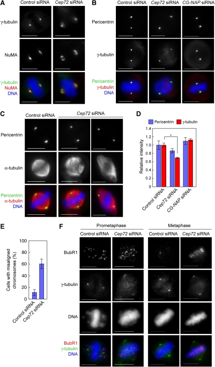
Figure 6.
Cep72-mediated attachment of spindle microtubules and mitotic centrosomes is required for proper chromosome alignment and segregation. (A) Control or Cep72-depleted cells were fixed and stained for NuMA, γ-tubulin, and DNA. (B) Control, Cep72, or CG-NAP siRNA-transfected cells were incubated in cold media containing 1 μM nocodazole for 30 min to depolymerize microtubules. Cells were fixed and immunostained for pericentrin, γ-tubulin, and DNA. (C) Microtubules were depolymerized as in (B). Mitotic cells were transferred to warm media to allow microtubules to re-grow. After recovering for 30 min, the cells were fixed and immunostained for pericentrin, α-tubulin, and DNA. Pericentrin staining indicates the position of the centrosomes. (D) Quantification of the relative intensity of γ-tubulin labelling at the centrosome in microtubule-depolymerized mitotic cells. Centrosomal areas were encircled by the edge of pericentrin staining and the mean intensities of pericentrin and γ-tubulin in each area were measured. Data show the average intensity in the centrosomal area and represent the mean±s.d. (n=20). *P<0.01. (E) The proportion of cells with misaligned chromosomes in control or Cep72-depleted cells with a bipolar spindle. Data represent the mean±s.d. of four experiments (n>20 for each experiment). (F) Control or Cep72 siRNA transfected HeLa cells were immunostained with BubR1, γ-tubulin, and DNA. Scale bar is 10 μm.

