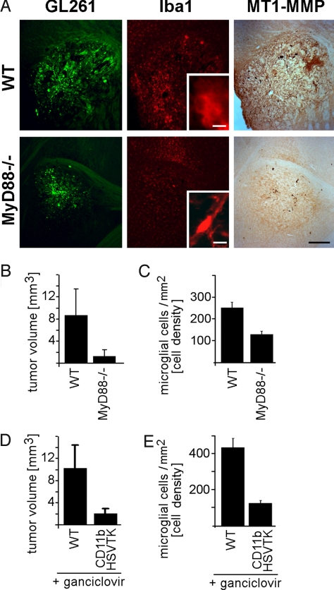
Fig. 5.
Glioma-induced TLR signaling in microglia promotes parenchymal MT1-MMP expression and tumor expansion in vivo. Mice with reduced TLR-signaling (MyD88-/-; n = 8) and WT controls (n = 8) were intracerebrally inoculated with GFP-expressing glioma cells (GL261, green) and, 14 d later, immunohistochemically analyzed for Iba1 (red) MT1-MMP (brown DAB precipitate) staining (A); note that tumor size, intra- and peri-tumoral microglia density, and labeling intensity for MT1-MMP are all reduced in MyD88-/- animals; tumor volume (B) and microglia density (C) in and around glioma from MyD88-/- and WT were also quantified by stereology. Glioma cells were injected into the brain of transgenic CD11b-HSVTK (n = 8) and WT (n = 8) mice with subsequent intratumoral microglia depletion by intracerebral ganciclovir infusion in transgenic mice. Tumor volume (D) and intra- as well as peri-tumoral microglia density (E) were quantified by a morphometric stereologic analysis; note the significant microglia depletion and reduced tumor size. (Scale bar: 500 μm in A, 10 μm in A Insets.)

