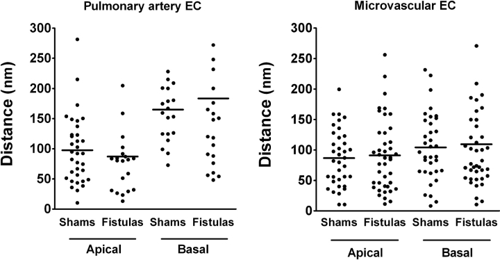
Figure 4.
Distances between the endoplasmic reticulum and the endothelial plasmalemmal membrane in intact lungs from shams (n = 4) and fistulas (n = 3). Data points and horizontal lines represent individual and average measurements, respectively, in pulmonary artery (left) and septal microvessel (right) endothelial cells (EC). Although the endoplasmic reticulum and the plasma membrane tended to be more distant from each other at the basolateral aspect compared with that in the apical compartment of pulmonary artery endothelium, this trend was not significant. We found no significant differences between shams and fistulas at either site in either cell population.

