
Figure 2.
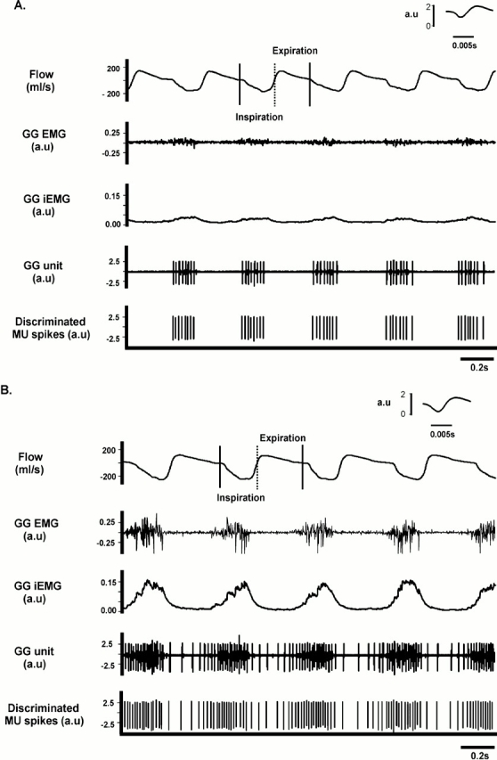
Original recordings of (from top down) airflow (with inspiration represented by a downward deflection), nonprocessed genioglossus EMG activity, the integrated EMG (iEMG) of the genioglossus, the nonprocessed genioglossus motor unit recording, and the discriminated motor unit spikes. (A) A segment of a recording obtained under baseline conditions; (B) recordings obtained during hypercapnia (inspired CO2 = 9%) in the same animal. Note that hypercapnia increased airflow and genioglossus EMG activity, as well as the frequency and train length of the active motor unit. Also note that the pattern of motor unit bursts transitioned from an inspiratory-only pattern under baseline conditions (A), to a preinspiratory–inspiratory pattern with hypercapnia (B). The insets shown in A and B represent a single spike recorded at high speed. See text for further details. MU = motor unit.