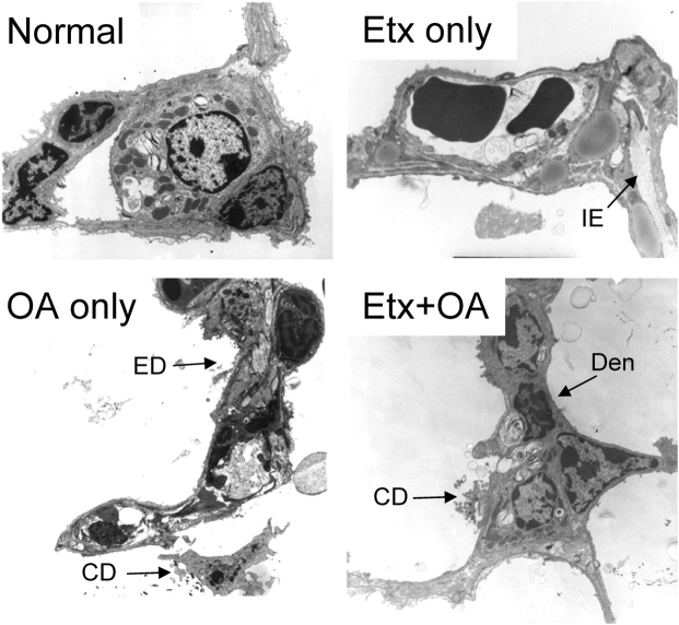
Figure 5.
Electron micrographs from all four experimental groups. The alveolocapillary membranes in the panels from normal and Etx-only mice are normal, although mild interstitial edema (IE) is present in the Etx-only panel. In contrast, there is widespread epithelial membrane damage (ED) and denudation (Den), fibrin and other cellular debris (CD), and proteinaceous edema in the alveolar space in the panels from mice given OA. Original magnification, ×3,000.

