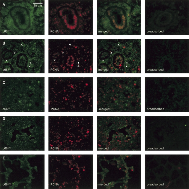
Figure 2.
Epithelial p66Shc and proliferating cell nuclear antigen (PCNA) expression both dissipate with maturation of the fetal baboon lung. By immunohistochemistry, p66Shc (green) was ubiquitously expressed in the 60-day lung (A). PCNA (red) was sparse in mesenchymal cells but widespread among epithelial cells. By 90 days, p66Shc was reduced and restricted to peribronchiolar mesenchymal cells and the apical cytoplasm of epithelial cells (B). PCNA expression persisted in most epithelial cells and increased in mesenchymal cells. Mesenchymal cells with high p66Shc content (arrowheads) tended to express less PCNA. Between 125 (C) and 175 (D) days, p66Shc expression shifted from isolated clusters of mesenchymal cells to discontinuous epithelial areas. Lungs from 140- and 160-day fetuses yielded variable and intermediate phenotypes in which p66Shc is expressed in clusters of both epithelial and mesenchymal cells. During this period, cells expressing PCNA continued to be scattered throughout the lung. The 3-day postnatal lung (E) expressed little p66Shc, and isolated cells expressing PCNA were scattered throughout the lung. Otherwise identically processed sections probed with preadsorbed p66Shc antibody yielded minimal fluorescence.

