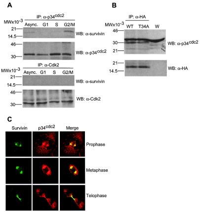
Figure 3.
Physical association between survivin and p34cdc2. (A) Cdk immunoprecipitation. HeLa cells asynchronously growing (Async.) or synchronized to G1, S, or G2/M were detergent-solubilized and immunoprecipitated (IP) with antibodies to p34cdc2 (Upper), or Cdk2 (Lower) followed by Western blotting with antibodies to survivin, p34cdc2, or Cdk2. (B) Survivin immunoprecipitation. HeLa cells transfected with HA-survivin or HA-survivin(T34A) were immunoprecipitated with an antibody to HA followed by Western blotting with antibodies to p34cdc2 or HA. W, whole extract. (C) Colocalization of survivin and p34cdc2 to the mitotic apparatus. Untransfected HeLa cells at the indicated phases of mitosis were labeled with mAb 8E2 to survivin (FITC, green) and a rabbit antibody to p34cdc2 (TR, red), and analyzed by confocal microscopy. Image merging analysis is shown on the right. (A and B) Relative molecular weight markers are on the left. WB, Western blotting.

