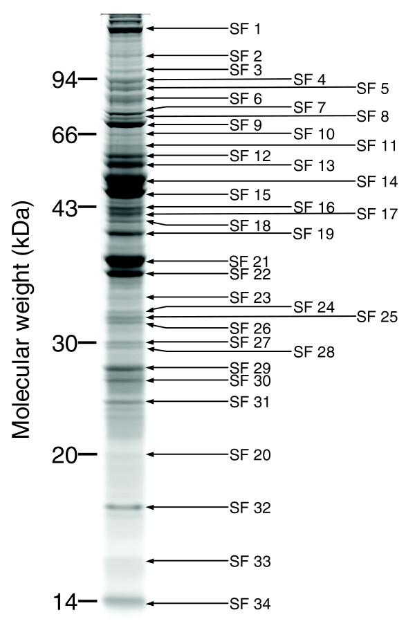
Figure 1.
SDS-PAGE gel separation of spermathecal fluid proteins. A colloidal Coomassie blue stained gel showing a representative protein profile of spermathecal fluid. A total of 50 μl of spermathecal fluid (SF) extract was loaded on the gel. Thirty-four protein bands, as indicated by arrows, were excised for protein identification. An overview of significant protein identifications for these bands is given in Additional data file 1.

