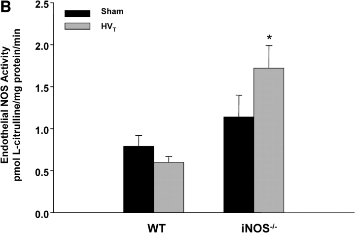
Figure 6.
Changes in lung NOS activity in response to MV. (A) iNOS activity. There was no detectable iNOS activity in lungs of iNOS−/− mice breathing spontaneously (controls) or exposed to HVT. In contrast, there was minimal iNOS activity in control WT mice, with a significant increase in response to HVT (n ⩾ 4 for each condition). *p < 0.05 versus control animals. (B) eNOS activity. At baseline (spontaneous breathing), lungs of both WT and iNOS−/− mice displayed activity. HVT resulted in a mild decrease and a significant increase in that activity in WT and iNOS−/− mice, respectively. *p < 0.05 versus control animals.


