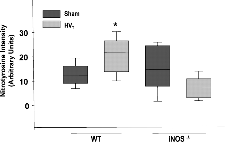
Figure 9.
Assessment of nitrotyrosine immunostaining in lung parenchyma of C57BL/6J (WT) and iNOS−/− mice exposed to HVT. Average was obtained through digital imaging of at least 20 fields/group (Nikon Eclipse E800 microscope with Pro+4.51 software; Nikon Instech Co., Kanagawa, Japan). *p < 0.05 versus sham.

