
Figure 4.
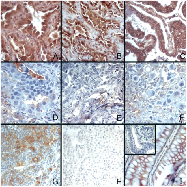

Top: TβRII immunostaining is decreased in invasive lung adenocarcinoma tumors. TβRII is expressed in BAC tumor cells (2+ immunoreactivity), localized to the cytoplasmic membrane and cytoplasm, with less intense staining detected in macrophages and endothelial cells (A–C); immunoreactivity is decreased or absent in well (D), moderately (E), and poorly (F) differentiated lung adenocarcinoma tumors. (G) Pituitary adenoma with 2+ immunoreactivity for TβRII (positive control). (H) Pituitary adenoma with isotype-matched, concentration-matched antibody showing no immunoreactivity (nonimmune antibody control). (I) Colonic mucosa showing 2+ immunoreactivity for TβRII, and inset showing invasive colonic adenocarcinoma from same section with no immunoreactivity for TβRII. Hematoxylin–eosin stain; original magnification, ×150. Bottom: TβRII immunostaining was highest in tumors with the least invasion. TβRII intensity: white bars = 0; gray bars = 1; black bars = 2. Slides of 55 independently acquired primary lung adenocarcinoma specimens were reviewed. The greatest linear dimension of histologic invasiveness was measured, which was subset into tertiles. The Spearman correlation coefficient of the relation between staining intensity and length of invasion was −0.36, p = 0.007.