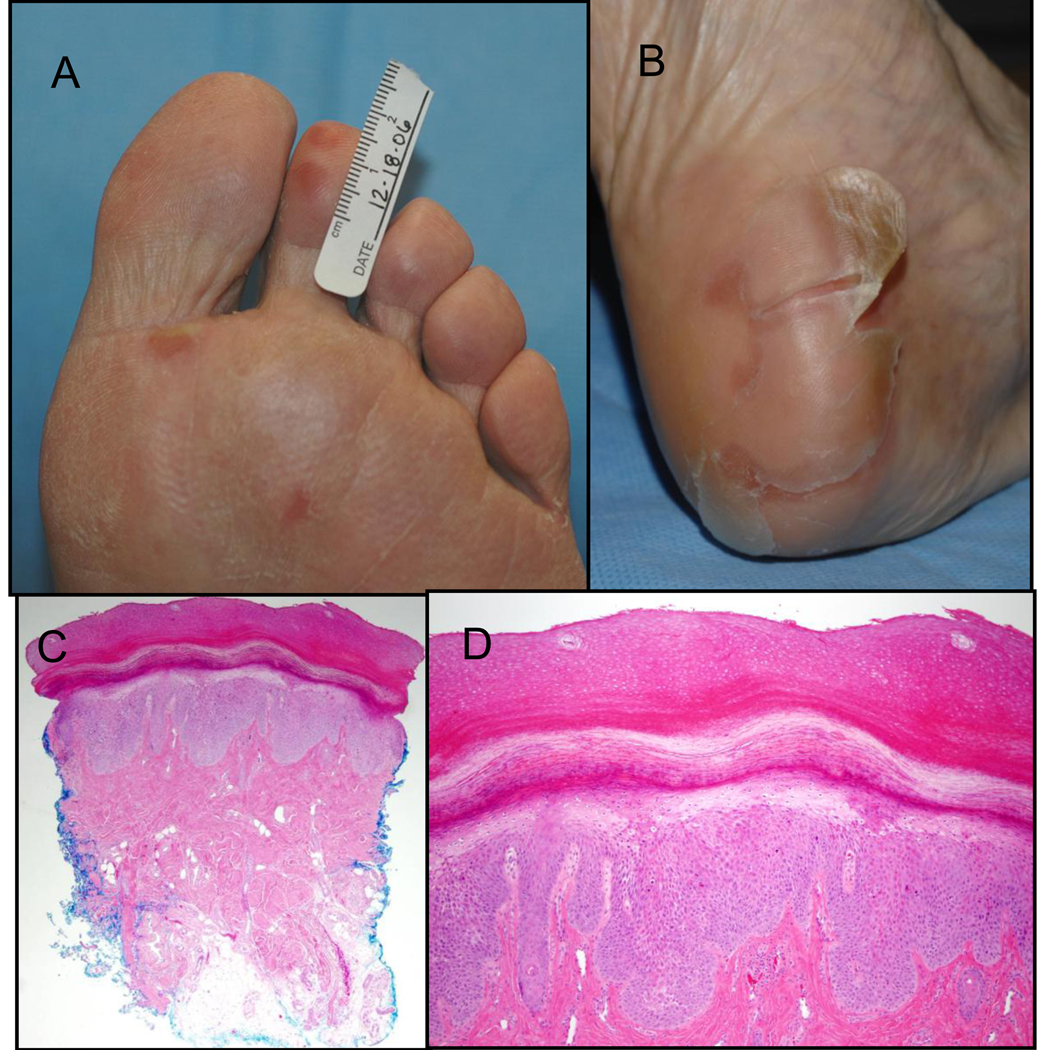
Figure 1.
A) Grade 2 hand-foot skin reaction showing early tender erythematous plaques at pressure points; B) Grade 2 hand-foot skin reaction demonstrating large sheets of desquamating skin overlying a tender erythematous plaque on the heel; C) Histology of an early HFSR lesion from Figure 1A shows epidermal thickening, reactive epithelial changes in the basal layer of the epidermis and in eccrine sweat ducts, mild perivascular infiltrate, and mild vascular dilatation (40X); D) Higher magnification of HFSR histology from Figure 1A (100X)

