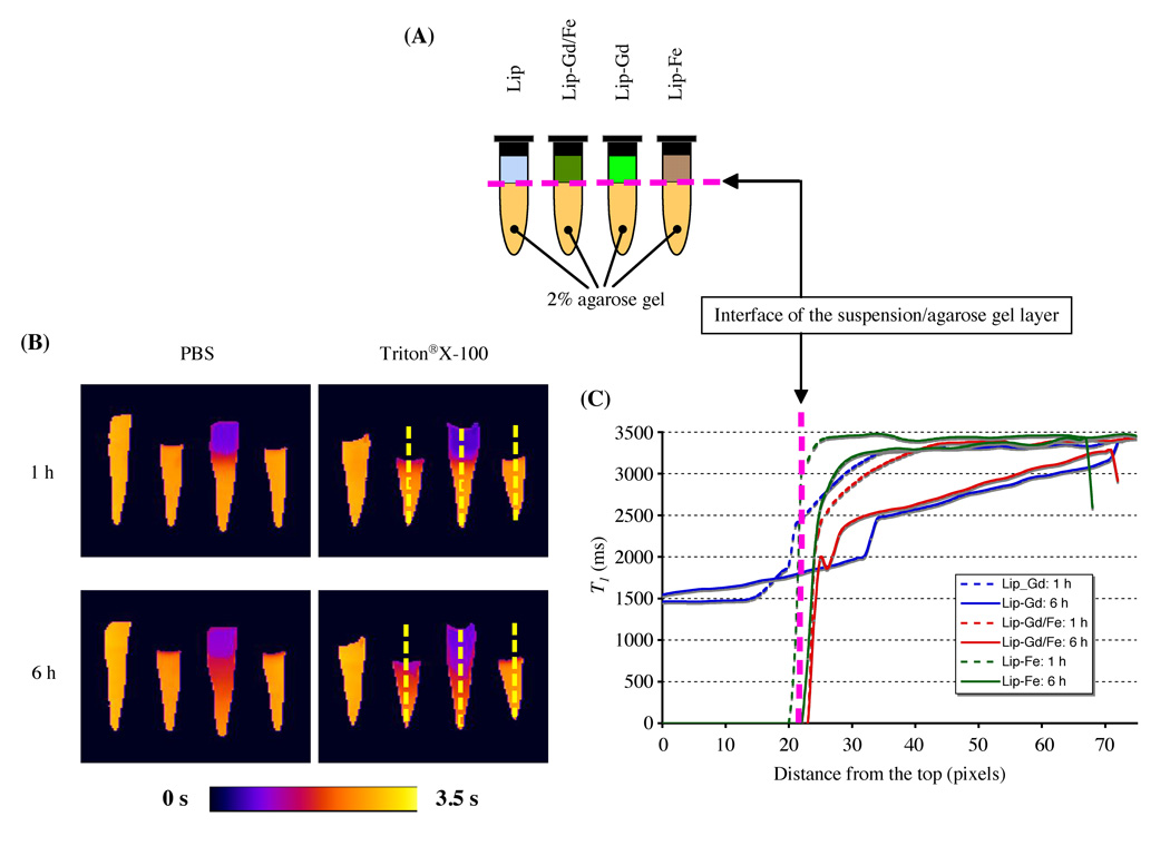
FIG. 2.
Visualization of the in vitro release of Gd-based contrast agent by the dual MR contrast agent technique. (A) Schematic of liposome phantoms. A mixture of 40 µL of liposome samples and 40 µL of either PBS (pH 7.4) or 10 mM Triton® X-100 were placed on the 2% agarose gel layer. (B) Quantitative T1 maps of liposome phantoms. Parameters for MRI are as follows: field of view = 40 × 22 mm; matrix size = 128 × 80; echo time = 15 ms; number of acquisitions = 2; three sagittal slices (slice thickness = 1 mm). (C) Diffusion profiles are expressed as a T1 value as a function of distance from the interface. Diffusion profiles of Lip-Gd/Fe in a 2% agarose gel layer were comparable to that of the free Gd-based contrast agent released from Lip-Gd.

