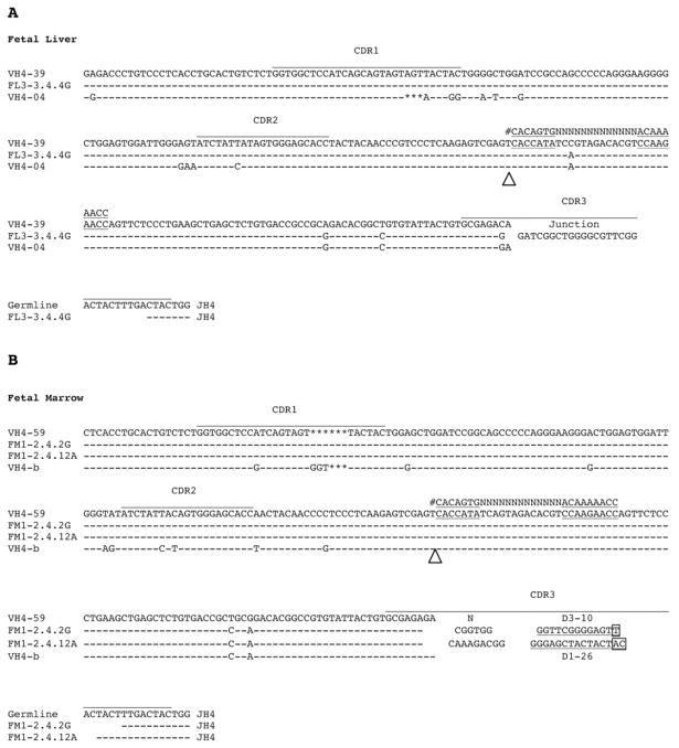
Figure 1. VH compound rearrangements from immature fetal and mature B cells.
Three examples of compound rearrangements with pseudohybrid joins found in fetal VH rearrangements are shown. The white arrowheads mark the proposed junction between the two VH genes. At the junction of the two VH genes, cRSS (heptamer-13 bp spacer-nonamer) which matches the canonical RSS is underlined. The canonical RSS is shown (#) for comparison. Asterisks mark deletional differences between the two germline genes (IGHV4-04/IGHV4-39 and IGHV4-b/IGHV4-59). (A) A compound rearrangement (VH FL3-3.4.4G) isolated from 18 week fetal liver CD19+IgM− B cells. The hybrid molecule consists of IGHV4-39 upstream of the cRSS and IGHV4-04 downstream. (B) Two VH compound rearrangements (FM1-2.4.2G, FM1-2.4.12A) isolated from 18 week fetal bone marrow CD19+IgM+ B cells. Both consist of IGHV4-59 upstream of the cRSS and IGHV4-b downstream.

