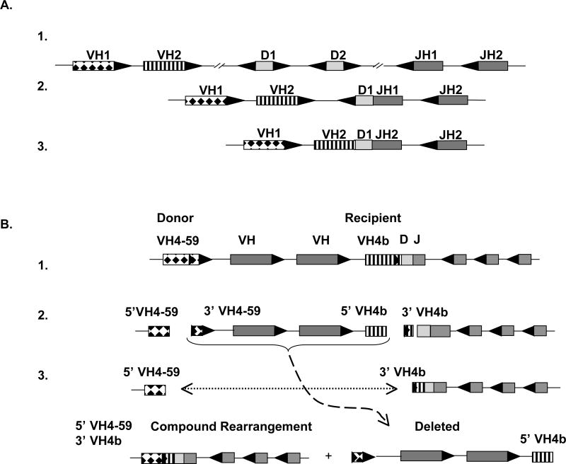
Figure 5. Schematic representation of a primary 12/23 VHDJH recombination and a secondary 12/12 rearrangement between two VH4 genes.
A. The heavy chain locus on chromosome 14 is partially represented to demonstrate normal VHDJH recombination. (1) The 23 RSS (black triangles) flank VH and JH genes and the 12RSS (white triangle) flank D segments. Each RSS which is targeted and cleaved by RAG1/2 is oriented with the heptamer adjacent to either the 5’ (D and JH) or 3’ (D and VH) end of the gene. (2) The D1 gene is joined to JH1 and the intervening D2 segment is deleted. (3) The VH1 gene is joined to the DJH segment and the intervening VH2 gene is deleted. B. A region of the heavy chain locus containing a primary IGHV4-bDJH rearrangement is represented. (1) RAG enzymes bind to the cRSS embedded in a non-rearranged VH4-59 donor gene and the cRSS of the recipient IGHV4-b gene that has undergone prior rearrangement with a D and JH segment. (2) RAG cleaves at the heptamer of the cRSS in IGHV4-59 and IGHV4-b (noted with arrows) (3) After RAG-mediated double strand cleavage at the cRSS heptamers, the intervening gene segment is removed and the 5’ portion of the donor gene (coding end) and the recipient VH4 gene containing the cRSS (signal end) are joined together forming a VH pseudohybrid join. Black triangles represent 23 bp RSS; white triangles represent 12bp RSS.

