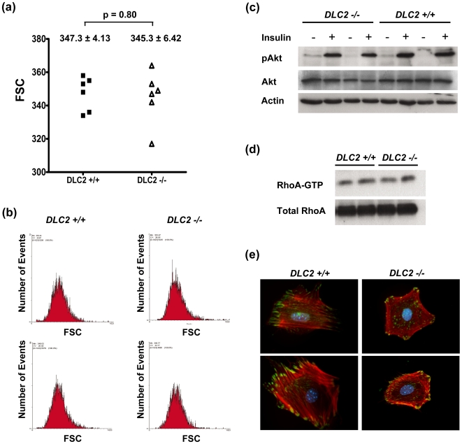
Figure 4. Depletion of DLC2 does not affect cell sizes.
(a) Comparison of sizes of DLC2+/+ (n = 6) and DLC2−/− (n = 6) cells by flow cytometry. Forward light scatter (FSC) of G1-phase-cells as determined by flow cytometry was used to measure the relative cell sizes. Mean values±standard errors and p-values are shown. P-values were calculated by unpaired Student's t test. (b) Representative samples of flow cytometry results. (c) Western blot analysis of insulin-treated DLC2+/+ and DLC2−/− cells. (d) RhoA activity of DLC2+/+ and DLC2−/− cells as detected by Rhotekin binding assay. (e) Actin cytoskeleton and focal adhesions in DLC2+/+ and DLC2−/− MEF cells were detected by immunofluorescence staining of actin stress fibers (red) and Paxillin (green), respectively.

