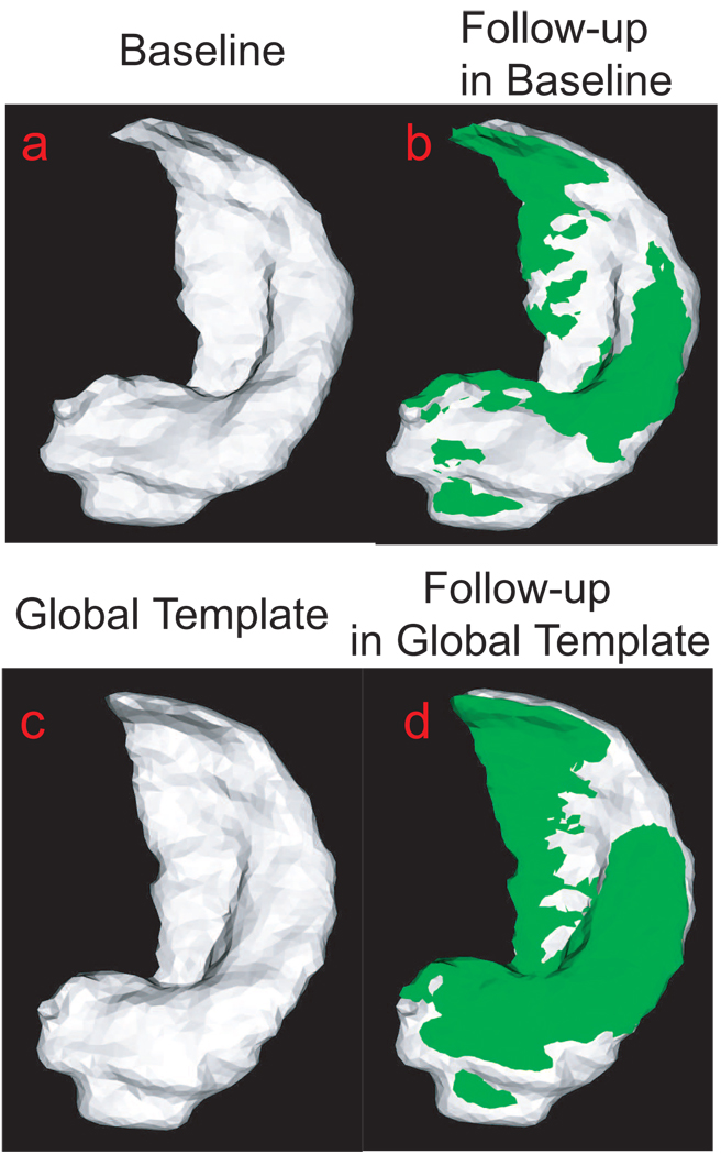
FIG. 6.
Panel (a) shows the hippocampus of a subject at the baseline. The hippocampus surface of this subject at the follow-up (green) is superimposed with one at the baseline in panel (b). Panel (c) shows the global hippocampus template. Panel (d) shows the hippocampus of this subject at the follow-up (green) represented in the global template coordinates (gray).

