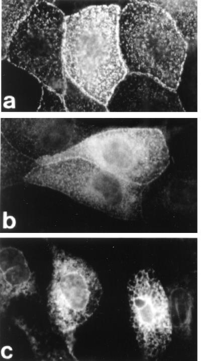
Figure 5.
Immunofluorescence of c-myc-tagged hSlo and the truncated mutant hSlo-S0–S6. Permanently transfected MDCK cells were grown on coverslips, treated with 12 mM sodium butyrate for 48 h, and fixed with 4% paraformaldehyde. Nonpermeabilized cells (a) and cells permeabilized with 0.2% Triton X-100 (b and c) were subjected to indirect immunofluorescence staining with anti-c-myc monoclonal antibody. hSlo shows a predominantly apical distribution (a and b), with a scant immune reaction inside the cell. Instead, the truncated mutant bearing a deletion after S6 (hSlo-S0–S6) showed no staining at the cell surface in nonpermeabilized cells (not shown), but revealed an intense intracellular reticular pattern characteristic of the ER in permeabilized cells (c).

