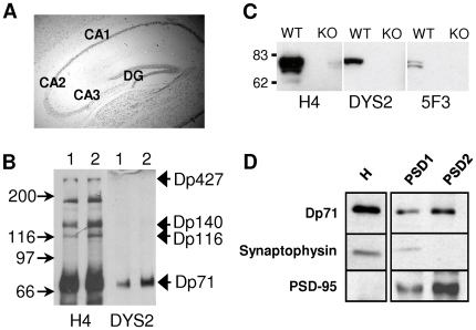
Figure 1. Regional and subcellular expression of Dp71 in brain.
A. Hippocampal Dp71 promoter activity in CA1-3 and dentate gyrus (DG) revealed by X-gal staining in brain section from a Dp71-null mouse. B. Immunoblots showing dystrophin-gene products detected by polyclonal (H4) and monoclonal (DYS2) antibodies in dissected CA (1) and DG (2) regions of the rat hippocampal formation. Note that in addition to Dp71, the H4 antibody also revealed Dp427, Dp140 and Dp116. Molecular weight markers are indicated on the left. C. Immunoblots of hippocampal extracts from WT and Dp71-null (KO) mice. Dp71 isoforms bearing exon 78 were detected by H4 and DYS2 antibodies, and those lacking exon 78 by 5F3. The doublet of bands detected by 5F3 likely reflects presence of glycosylated and non-glycosylated forms of Dp71f [5], [21]. D. Detection of Dp71 in the postsynaptic densities. Protein extracts from subcellular fractions obtained from control mouse brains were probed with the anti-Dp71 (H4) antibody (top panel) and with the presynaptic and postsynaptic markers synaptophysin (central panel) and PSD-95 (bottom panel), respectively. H: total homogenate; PSD1/PSD2: isolated PSD fractions (Detailed distribution in subcellular fractions is shown in Fig. S3).

