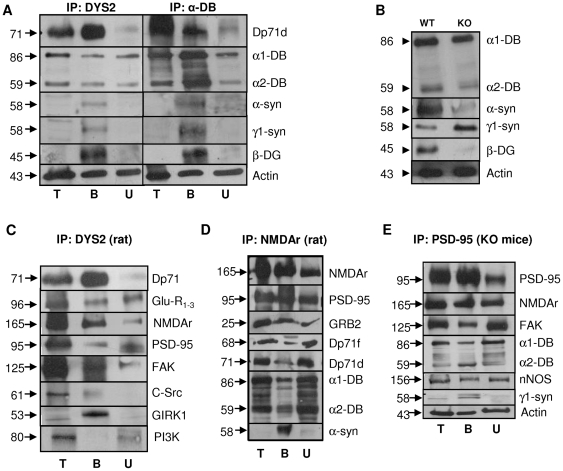
Figure 3. Characterization of the brain Dp71-DAPC.
A. Immunoprecipitation of Dp71 with DYS2 or of αDB with αDB antibodies in rat whole-hippocampus extracts. Co-immunoprecipitated proteins were analyzed on western blots. B. Immunoblots of hippocampal extracts from WT and KO mice using antibodies directed against α-DB, α-syn, γ1-syn, β-DG and actin. C. Immunoprecipitation of Dp71 with DYS2 and westernblot analysis of interacting proteins in rat hippocampus. Co-immunoprecipitated proteins were AMPAr subunits Glu-R1-3, NMDAr subunits NR2A-B, PSD-95, FAK, C-Src and GIRK1, not PI3K. D. Immunoprecipitation using the NMDAR2A&B antibody in rat hippocampal extracts. The proteins interacting with the NMDAr complex were PSD-95, GRB2, Dp71f and Dp71d, α-DB and α-syn. E. Immunoprecipitation of the PSD-95 protein from hippocampal extracts of KO mice; NMDAr, FAK, α-DB, nNOS and γ1-syn were pulled down in the bound fraction. α-DB: α-dystrobrevins, α-syn: α-syntrophin, γ1-syn: γ1-syntrophin, β-DG: β-dystroglycan. T: Hippocampus total homogenate; B: Bound fraction; U: Unbound fraction. Proteins molecular mass (left) are indicated on the right.

