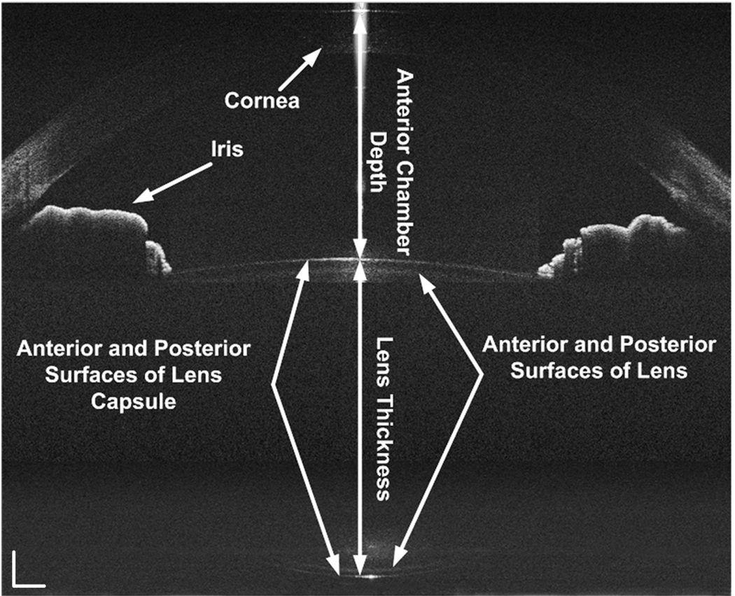
Fig. 3.
The simultaneously acquired images were constructed in scale, but with optical correction. All the surfaces of the anterior segment of the eye including the cornea, anterior chamber, anterior and posterior surfaces of crystalline lens and capsule were clearly visualized. The white bar: 0.5mm.

