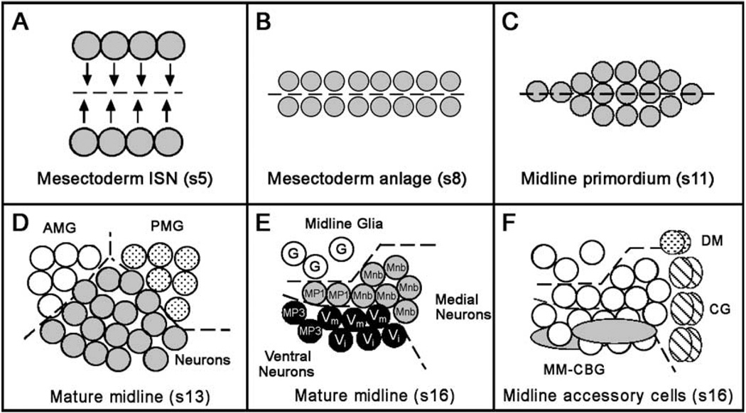
Fig. 1.
Schematic summary of CNS midline cell development. In all panels, a single segment is shown with anterior to the left. Embryonic stages are indicated by “s#”. (A) Mesectoderm ISN stage, ventral view. Two stripes of mesectodermal cells reside on either side of the mesoderm in the blastoderm embryo (stage 5). Dotted line indicates ventral midline of embryo. There are four cells/segment on each side. Arrows represent how the mesectodermal cells move together at the ventral midline during gastrulation (stage 6) as the mesoderm invaginates. (B) Mesectoderm anlage stage, ventral view. During the mesectoderm anlage stage (stages 7–8), the mesectodermal cells meet at the midline and then undergo a synchronous cell division, resulting in 16 cells per segment. (C) Midline primordium stage, ventral view. During the midline primordium stage, midline cells rearrange from a two-cell wide planar array into a cell cluster. Midline cells within these clusters differ slightly in their dorsal/ventral positions. (D) Mature CNS midline cells, stage 13. Sagittal view, dorsal up. At stage 13, two populations of midline glial cells become evident. The anterior midline glia (AMG; open circles) are reduced by apoptosis but ultimately will ensheathe the commissures while all posterior midline glia (PMG; dotted circles) will undergo apoptosis. Midline neurons (shaded circles) occupy the space between and below the midline glia. Dotted lines separate the different cell groups. (E) Mature CNS midline cells, stage 16. Sagittal view, dorsal is up. The PMG have undergone apoptosis and are absent, whereas the AMG give rise to ~3 mature glia (G, open circles). Midline neurons have migrated to their final positions within the ganglion. Medial neurons include MP1 neurons (MP1, shaded circles) and the progeny of the MNB (Mnb, shaded circles). Ventral neurons include VUM motorneurons (Vm, black circles), VUM interneurons (Vi, black circles), and MP3 neurons (MP3, black circles). (F) Midline accessory cells shown in relation to midline neurons and glia (open circles). Two DM cells (dotted circles) lie atop the CNS near the midline channel, which is lined by six-channel glia (CG; hatched ovals). The two MM-CBG in each segment (shaded ovals) are closely associated with the ventral neurons.

