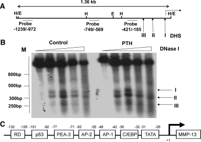
Figure 1.
Analysis of DNase I-hypersensitive sites in the rat MMP-13 promoter in rat osteoblastic cells. Nuclei isolated from rat osteoblastic osteosarcoma UMR 106-01 cells untreated (control) and treated with rat PTH (peptide 1–34, 10−8 m) for 30 min were digested with increasing amounts of DNase I (0, 20, 40, 80, and 160 U per 107 nuclei); the DNAs were purified and digested with HindIII or with EcoRI. Each sample (30 ug) was separated on 2% agarose gels and characterized by Southern blot analysis. A, Partial restriction map of rat MMP-13 promoter showing the hybridization probes and the positions of the DNase I-hypersensitive sites (DHS I, DHS II, DHS III) relative to the transcription start site. E, EcoRI; H, HindIII. B, Autoradiograms from hybridizations with [α32P-]dCTP-labeled probe −421/−185. The positions of the DNase I-hypersensitive sites (I, II, III) are marked with arrows. C, Schematic representation of the promoter of the rat MMP-13 gene. From right to left are sites for the following consensus sequences: C/EBP, AP-1, AP-2, PEA-3, p53, Runx2 distal. C/EBP, CCAAT enhancer-binding protein.

