
Figure 2. Wg signaling via Fz and DFz2 receptors is required for the proper maintenance of hemocyte precursors.
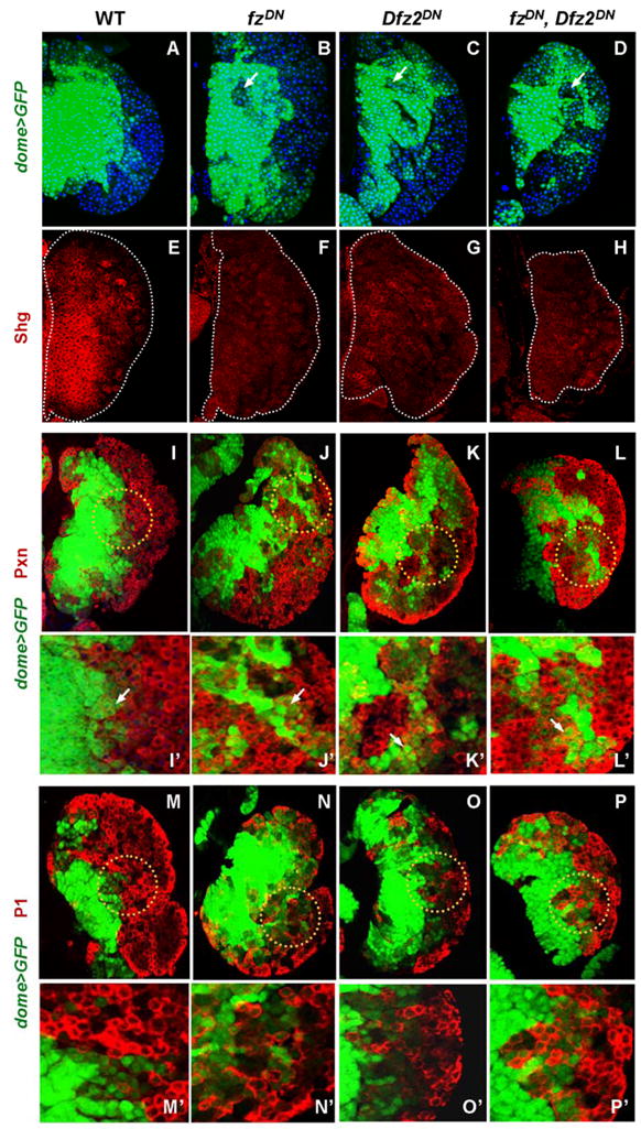
Genotypes (dome+ is an abbreviation for dome-Gal4, UAS-GFP) are listed on the top and drivers and antibodies for detection on the left edge of the panels. Antibodies and the markers are color coded in the appropriate panels. Images represent accrued 6μm confocal sections through the LG. (A–D) Redistribution of dome+ cells in fz/Dfz2 mutants. (A) In wild type (WT), prohemocytes marked by dome (green),are restricted to the medullary area of the LG.
In single mutant (B, C) and in double mutant (D) backgrounds, dome>GFP+ prohemocytes can be found in cortical areas while dome>GFP negative cells often reside inside the MZ compartment (indicated by arrows).
(E–H) Expression of DE-cadherin (Shg) is down regulated in fz/Dfz2 mutants. In WT (E) Shg (red) is expressed in the MZ, while in single (F, G) and double (H) mutants of the receptors, the expression of Shg is significantly reduced (edges of the LG are outlined by a dotted line).
(I–L′) Expression of Pxn, an early maturation marker,is up regulated in dome>GFP+ cells of fz/Dfz2 mutants. In wild type (I, I′), Pxn is expressed in mature hemocytes of the CZ and in rare dome+ hemocyte precursors located at the edge of the MZ (indicated by arrow in I′, which is the magnified area within the dotted circle in I). In single mutants (J, J′ and K, K′) and in the double mutant (L, L′) there is a significant increase in dome+/Pxn+ double positive cells that represent immature hemocytes in a transition state of differentiation (J′ to L′, indicated by arrows).
(M–P′) In fz/Dfz2 mutants the terminal differentiation of plasmatocytes is not affected as revealed by the expression of the late differentiation marker, P1. In WT (M, M′), as well as in single (N, N′ and O, O′) and in double (P, P′) mutants, P1 expression is restricted to mature hemocytes of the CZ. Higher magnification images (M′ to P′) of corresponding areas of LG outlined in M through P are shown. Lymph glands were analyzed at 68h after larval hatching maintained at 29°C.