
Figure 2.
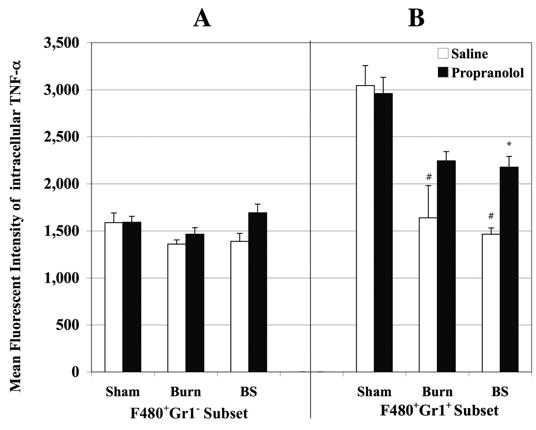
Bar graph delineating MFI of intracellular TNF-α produced by monocytes obtained from peripheral blood of sham (S), burn (B) and burn sepsis (BS) animals with saline (open bars) and propranolol (black bars) treatments. Panels A and B represent ic-TNF-α produced by F4/80+Gr1− resident monocytes and F4/80+Gr1+ inflammatory monocytes respectively. Burn and burn sepsis significantly reduced ic- TNF-α production by Gr1+F4/80+ monocyte subsets compared to sham (# p< 0.05 vs. S and B). Propranolol restored the inflammatory potential of Gr1+F4/80+ monocyte subsets in BS group (* p < 0.05 vs. saline treatment in BS). n= 4 in S and B, n=6 in BS.