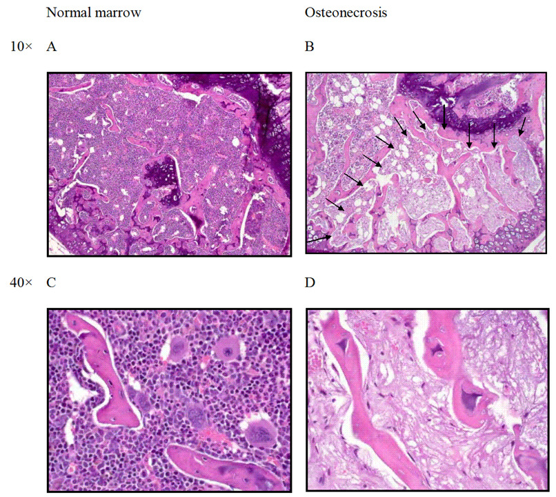
Figure 1.
Histologic features of osteonecrosis in a BALB/cJ mouse treated with oral dexamethasone (4 to 2 mg/L). (A,C) Normal architecture of the distal femoral epiphysis with densely cellular hematopoietic elements in the marrow and prominent osteocyte nuclei observed in trabecular bone. (B) Arrows outline segmental osteonecrosis in the distal femoral epiphysis. (D) Higher magnification of osteonecrotic lesion with necrotic marrow elements and empty osteocyte lacunae.

