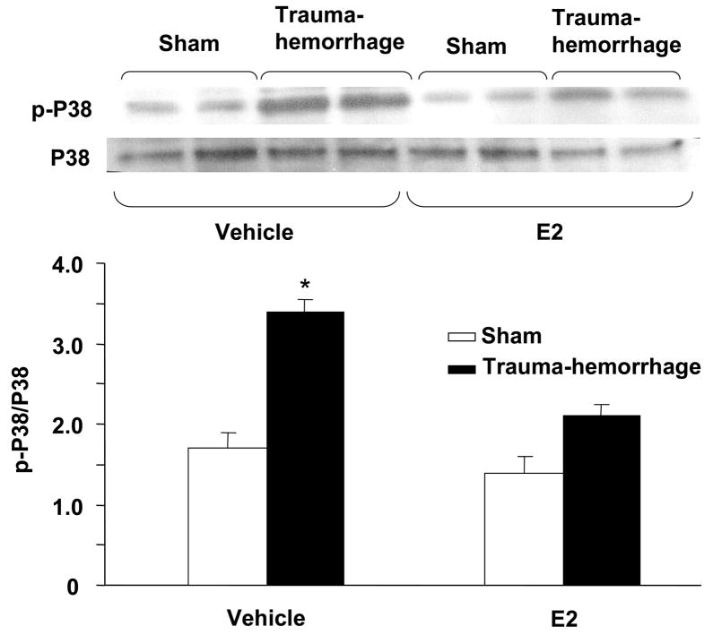
Figure 4.
p38 phosphorylation and protein expression in epidermal keratinocytes following trauma-hemorrhage. Keratinocytes were isolated (1×10⁶ cells/ml) and stimulated with LPS (5μg/ml) for 30 min and lysed, lysates were analyzed for p38 phosphorylation by Western blot. The blots were stripped and reprobed for p38 total protein contents in various lanes. Blots from 6 animals in each group were analyzed using densitometry. Densitometric values for phosphorylation were normalized to p38 protein and are shown in bar graph as mean ± SEM. *p<0.05 compared to all other groups.

