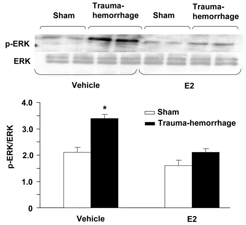
Figure 5.
ERK phosphorylation and protein expression in epidermal keratinocytes following trauma-hemorrhage. Keratinocytes were isolated (1×10⁶ cells/ml) and stimulated with LPS (5μg/ml) for 30 min and Lysates of keratinocytes were analyzed for ERK phosphorylation by Western blot. The blots were stripped and reprobed for ERK total protein contents in various lanes. Blots from 6 animals in each group were analyzed using densitometry. Densitometric values for phosphorylation were normalized to ERK protein and are shown in bar graph as mean ± SEM. *p<0.05 compared to all other groups.

