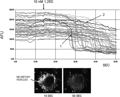
FIG. 1.
1,25D induces exocytotic release from quinacrine-loaded secretory vesicles in osteoblastic ROS 17/2.8 cells. Fluorescence intensity (expressed as arbitrary fluorescence units [AFUs]) recorded continuously from 45 individual secretory vesicles in a ROS 17/2.8 osteoblast during the addition of 10 nM 1,25D to the extracellular bath. Quinacrine-stained vesicles were selected from the image of an osteoblast shown in the bottom left panel. Continuous recording of fluorescence intensity was done over 52 s; 1,25D was added at the time indicated by the arrow, ∼14 s after the beginning of the recording. Nine of the selected areas are indicated with circles drawn on the image on the left, which depicts the osteoblast immediately before treatment with the 10 nM 1,25D, at time 10 s from the beginning of the recording. The image on the right shows the same osteoblast after exocytotic release of quinacrine, at the end of the recording. The average AFU intensity of each selected area was measured every 500 ms. Highlighted for comparative purposes are traces obtained for a vesicle undergoing complete exocytosis (labeled 1, intensity decrease from ∼100 to <50 AFU about 27 s into the recording), and a vesicle that did not undergo exocytosis (labeled 2, fluorescence intensity remained between 200 and 250 AFU during the entire length of the experiment).

