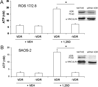
FIG. 5.
1,25D induction of exocytotic ATP release requires a classic VDR in osteoblasts. ATP concentration values (expressed in nM) measured in the extracellular bath 2 min after the addition of 10 nM 1,25D or 0.01% ethanol (vehicle control) to ROS 17/2.8 (A) and SAOS-2 (B) cell cultures expressing the native VDR vs. cultures in which the VDR was silenced (−VDR). (Right panels, top and bottom) Western blot analysis for VDR expression in native (control vector-transfected) and siRNA VDR-silenced osteoblasts; α-vinculin was used as a loading control. Data shown are mean values ± SE obtained from n = 5–6 independent experiments. *p < 0.05.

