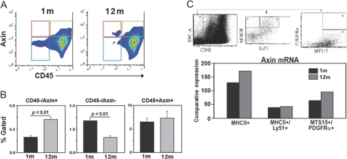
Fig. 2.
Axin expression in TSC populations. (A and B) The thymi from young (n=6) and 12-month-old (n=6) mice were digested enzymatically and labeled with Axin-AF-488 and CD45-APC. CD45–Axin+ cells (in red box) are increased significantly (P<0.05) in older mice, CD45–Axin– (blue box) decrease with age, and no change in CD45+Axin+ (green box) is detected. All data are expressed as mean ± sem. (C) The CD45 cells were depleted from thymic digests using magnetic bead-based positive selection. The CD45– cells from six thymi, each from 2- and 12-month-old mice were pooled and used for sorting the mTEC (CD45–MHCII+Ly51–), cTEC (CD45–MHC11+Ly61+), and fibroblasts (MTS15+PDGF-Rα+). The comparative threshold values from triplicate real-time PCR analyses were collapsed together, and data are expressed as relative axin mRNA expression. SSC-A, Side-scatter-A.

