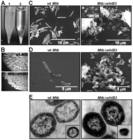
Figure 1. Mtb WhiB3 regulates cell shape and size.
(A) wt Mtb and MtbΔwhiB3 were cultured in 7H9 liquid media containing 0.1% Tween 80 till stationary phase. At an OD600 nm = 4.5 the aggregation phenotype was examined by allowing the cells to settle for 10 min. MtbΔwhiB3 cells settled rapidly whereas wt Mtb remained dispersed (B) Spot colony rugosity of wt Mtb and MtbΔwhiB3 was analyzed by spotting identical volumes (30 µl) and equal number of cells (3×10⁶) on Dubos complete medium. Cells were allowed to grow for 4 weeks and photographs were taken at 7× magnification using a Zeiss stereo microscope. (C and D) SEM analysis demonstrating that WhiB3 affects cell length. Low magnification SEM (C, inset) illustrates the severe clumping of MtbΔwhiB3 cells. Approximately 5 SEM fields (10 cells/field) were analyzed to determine the cell sizes of wt Mtb and MtbΔwhiB3. (E) TEM analysis showing hyperstaining of MtbΔwhiB3 cells as compared to wt Mtb cells. Inset; low magnification image.

