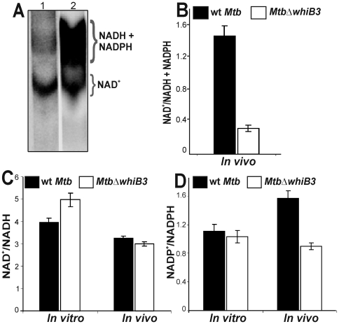
Figure 6. Mtb WhiB3 maintains the intrabacterial NAD+/NADH and NADP+/NADPH poise.
(A) TLC analysis of oxidized and reduced pyridine nucleotides labeled with [14C] nicotinamide. Wt Mtb (lane 1) and MtbΔwhiB3 (lane 2) cells isolated from infected macrophages (∼108 cells) were labeled with [14C] nicotinamide for 24 h in 7H9 basal medium containing acetate as a carbon source and analyzed by TLC by loading equal cpm in each lane. Note the strong labeling of reduced pyridine nucleotides from MtbΔwhiB3 isolated from macrophages. (B) Densitometric analysis of the relative abundance of nucleotides in (A). Experiments were performed four times and similar observations were recorded. Cells grown in vitro or isolated from the infected macrophages were analyzed by enzymatic assays using (C) alcohol dehydrogenase for NAD+/NADH estimation and (D) glucose-6-phosphate dehydrogenase for NADP+/NADPH analysis. Data shown is the average of two independent experiments.

