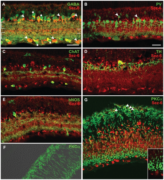
Figure 3. Immunostaining for Sez-6 and retinal subtype markers.
Rat retina vertical sections double stained for Sez-6 (red) and (in green): (A) GABA – non-GABAergic amacrine and putative displaced amacrine cells are marked (arrowheads) while double-labelled amacrine cells are marked with asterisks. (B) Parvalbumin (PV) – Sez-6 positive amacrine cells with their somata located in the second and third layer from the IPL (arrowheads) and double-stained cells with the elongated soma typical of bipolar cells (arrows) are observed. (C) Choline acetyltransferase (ChAT)-positive amacrines in the INL and displaced amacrines in the GCL were double-labelled with Sez-6. (D) Dopaminergic amacrine cells (labelled with tyrosine hydroxylase) were rarely found however they co-labelled for Sez-6. (E) Brain nitric oxide synthase (bNOS) amacrine cells were also positive for Sez6 staining. (F & G) Immunoreactivity for Protein kinase C alpha (PKCα - green) in rod bipolar cells overlaid on a bright field image (F) or shown with Sez-6 in red (G) indicating Sez6 staining at the apical pole of the cell soma (arrowheads). Scale bars: 25 µm.

