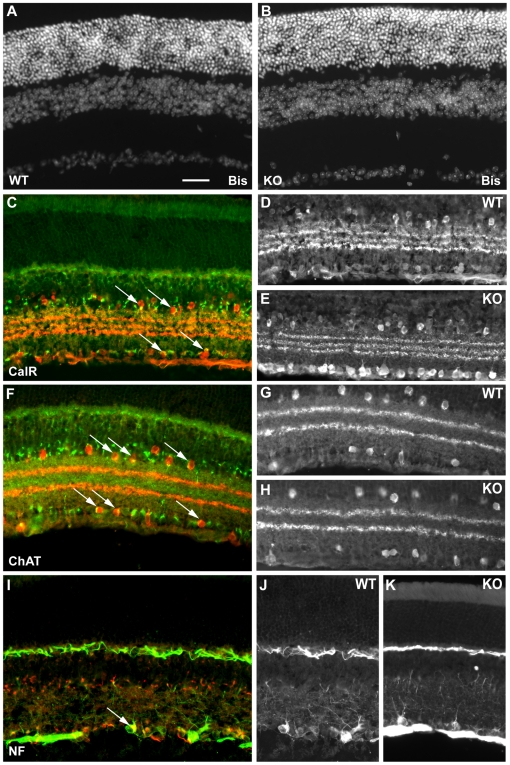
Figure 4. Immunostaining of Sez-6 wild-type and knockout mouse retina.
Vertical sections of Sez-6 WT (A) and KO (B) mouse retina stained with bisbenzamide (Bis) reveal no obvious morphological differences between the genotypes. (C) Sez-6 WT retina stained for calretinin (CalR – red) and Sez-6 (green) showing double stained amacrine and displaced amacrine cells in the INL and GCL (arrows). (D) Sez-6 WT and (E) KO sections from the central retina stained for CalR displaying a typical staining pattern. (F) Sez-6 WT retina stained for ChAT (red) and Sez-6 (green). Double-labelled cells in the INL and GCL are indicated (arrows). (G) Sez-6 WT and (H) KO retina stained for ChAT alone show an identical staining profile. (I) Sez-6 WT retina stained for neurofilament (NF - green) and Sez-6 (red). A double-stained cell morphologically similar to an alpha (or A1) ganglion cell is marked (arrow). (J) Sez-6 WT and (K) KO retinal sections stained for neurofilament alone showing a similar density of labelled cells in the GCL in both genotypes. Scale bar = 25 µm.

