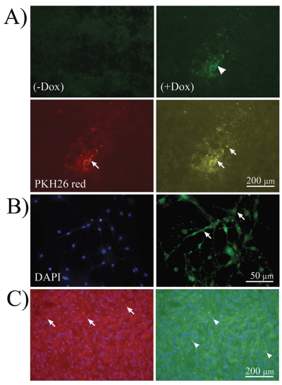
Fig. 2.
In vivo ESC-mediated conditional TRAIL expression. (A) Immunofluorescence microphotographs of A172 tumor xenografts injected with embryonic stem cell (ESC)-derived astrocytes conditionally expressing TRAIL after immunohistochemistry for TRAIL antibody in the absence of doxycycline (− Dox; control) or presence of Dox (+Dox). TRAIL expression was present in experimental animals (arrowhead, upper right) and not in control. TRAIL protein was visualized by Alexa 488 antibody. Lower left, ESC-derived astrocytes tracked with PKH26 vital red (arrow; 565-wavelength filter). Lower right, Computer-overlapped images of PKH26-tracked ESC-derived astrocytes and TRAIL immunolabeling. Note the colocalization (yellow) of both within the same cells (arrows). (B) At higher magnification, ESC-derived astrocytes expressing TRAIL show membrane localization of the protein (arrows). Left, 4’,6-diamidino-2-phenylindole (DAPI) staining; right, TRAIL immunolabeling. (C) “Homing” after systemic injection was seen in experimental animals. Left, PKH26-labeled astrocytes are shown as red punctuate labeling embedded in tumor cells (arrows). Right, astrocytes expressing TRAIL in green embedded within the tumor (arrowheads).

