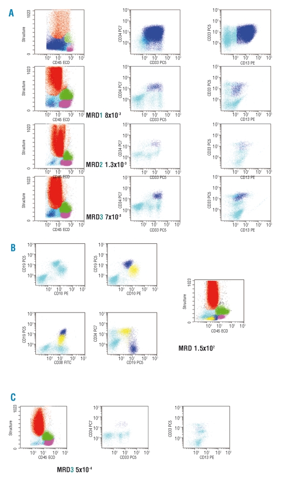
Figure 2.
Monitoring minimal residual disease (MRD) in FCM. (A) Backgating of leukemic cells on a CD45/SSC flow cytometry scatter-gram. The procedure of backgating was used to superimpose the population of cells identified at diagnosis and as MRD by multiparameter flow cytometry in bone marrow samples of a patient with AML. Color legend:granulocytes in red, monocytes in green, lymphocytes in purple, immature cells in cyan, MRD in dark blue. (B) Hematogones and B-ALL leukemic cells. Hematogones are shown in dark blue while the remaining blasts are displayed in yellow. Other colour code as above. (C) MRD in peripheral blood of an AML patient. Same patient as in panel A.

