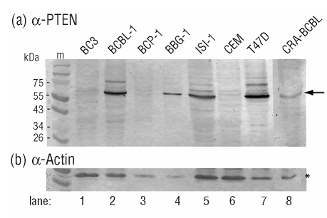
Figure 1.
PTEN expression in PEL cell lines. Lysates from BC-3 (lane 1), BCBL-1 (lane 2), BCP-1 (lane 3), BBG-1 (lane 4), ISI-1 (lane 5) and CRA-BCBL (lane 8) cell lines were analyzed by Western blot for PTEN (A) and actin (B) protein expression. The T-ALL CEM (lane 6) and breast adenocarcinoma T47D (lane 7) cell lines served as negative and positive controls, respectively. Sodium dodecyl sulfate (SDS)-denatured cellular proteins were separated by SDS-polyacrylamide gel electrophoresis (PAGE) in 10% acrylamide gels using a discontinuous buffer system (Laemmli, U.K., 1970). The transfer of proteins onto nitrocellulose membranes (Hybond ECL, Amersham Biosciences) was carried out using a semi-dry blotting system. Blots were blocked with skimmed milk in TNT buffer (20 mM Tris-HCl pH 7.5, 150 mM NaCl, 0.05% Tween-20) and developed after successive incubation with anti-human PTEN rabbit antibody (R&D Systems) and alkaline phosphatase-labeled goat anti-rabbit IgG conjugate (Sigma-Aldrich).

