Figure 3.
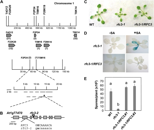
Map-based cloning of rfc3-1. A, Map of the rfc3-1 locus on chromosome 1. Positions of the markers used for mapping are indicated. The two final flanking markers were F2P24 and T5M16. The rfc3-1 mutation is marked with an asterisk. B, Gene structure of RFC3. Exons are indicated with boxes, and introns are represented by lines. The region where the G-to-A mutation occurs in rfc3-1 is shown in detail. C, Morphology of the wild type (WT), rfc3-1, and rfc3-1 transformed with a genomic clone of RFC3 driven by its native promoter. Plants were grown on soil for 4 weeks before the photograph was taken. D, pBGL2-GUS reporter gene expression in rfc3-1 and rfc3-1 transformed with a genomic clone of RFC3 driven by its native promoter. Two-week-old seedlings grown on MS with or without 10 μm SA were stained for GUS activity as described previously (Bowling et al., 1994). E, Growth of H. a. Noco2 on the wild type, rfc3-1, and two independent transgenic rfc3-1 lines carrying genomic RFC3 driven by its native promoter. The experiment was carried out as described in Figure 1E. Statistical differences among the samples are labeled with different letters (P < 0.01, ANOVA). The experiment was repeated at least three times with similar results.

