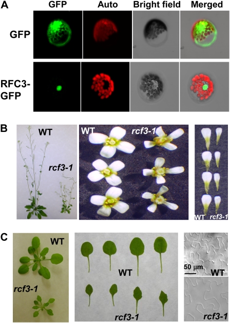
Figure 5.
Functional analysis of Arabidopsis RFC3. A, Localization of RFC3-GFP. Arabidopsis mesophyll protoplasts expressing the pG229-RFC3-GFP fusion proteins were examined by confocal microscopy, and the image shown was taken from a representative protoplast. The red fluorescence reflects chlorophyll autofluorescence. The nucleus is obvious from the bright-field image of the protoplast. pG229-GFP alone was used as a control. The experiment was repeated once with similar results. B, Morphology of mature plants, representative flowers, and petals of the wild type (WT) and rfc3-1. C, Morphology of young seedlings and leaves of the wild type and rfc3-1. The first to fourth true leaves of 20-d-old seedling were used to measure the epidermal cells with a microscope. The scale bar is the same for both images.

