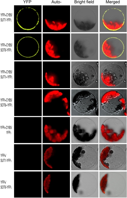
Figure 4.
In vivo interaction of MdCYB5 with MdSUT1 and MdSOT6 in the BiFC system. The laser-scanning confocal microscopy images show fluorescence (indicated by YFP) and merged images of the double transgenic cells with the YFPN-MdCYB5 and MdSUT1-YFPC fusions (YFPN-CYB5/SUT1-YFPC) or with the YFPN-CYB5/SOT6-YFPC construct pair. The wild-type MdSUT1 and MdSOT6 were replaced in the above-mentioned constructs by the mutated MdSUT1P (Leu-73→Pro; YFPN-CYB5/SUT1P-YFPC) and mutated MdSOT6P (Leu-117→Pro; YFPN-CYB5/SOT6P-YFPC), respectively, which were used to transform cells that served as negative controls. The construct pairs YFPN-CYB5/YFPC, YFPN/SUT1-YFPC, and YFPN/SOT6-YFPC were also used as negative controls to transform the protoplasts. The chlorophyll autofluorescence (Auto-) and the bright-field images are also presented.

