Figure 5.
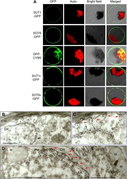
Localization of MdSUT1, MdSOT6, and MdCYB5 in cells. A, Transient expression of MdSUT1-GFP (and its mutated form MdSUT1P), MdSOT6-GFP (and its mutated form MdSOT6P), and MdCYB5-GFP fusion proteins in the Arabidopsis protoplasts. The laser-scanning confocal microscopy images show the MdSUT1 (indicated by SUT1-GFP), MdSOT6 (SOT6-GFP), GFP-MdCYB5 (GFP-CYB5), MdSUT1P (SUT1P-GFP), and MdSOT6P (SOT6P-GFP) fluorescence (GFP) and the merged images. The chlorophyll autofluorescence (Auto-) and the bright-field images are also presented. Note that the mutation of MdSUT1P did not change the plasma membrane localization of MdSUT1, and the mutation of MdSOT6P did not change the plasma membrane localization of MdSOT6. B to E, Immunogold labeling of MdCYB5 in apple fruit cells. The protein reacting with anti-MdCYB5N (visualized by immunogold particles) mainly resides on the ER (B and D). A blowup (C) of the area from B shows more clearly the ER localization of MdCYB5; note that some of the ER hosting MdCYB5 immunoparticles distribute near the plasma membrane (PM), which are shown by arrows. D shows more clearly the distribution of MdCYB5-hosting ER around the plasma membrane (indicated by arrows). No substantial signal was detected in other cellular compartments (B–D) such as the cell wall (CW), mitochondrion (M), and cytoplasm. No substantial gold particles were found in the control cells without antiserum treatment (E). Bars = 0.5 μm.

