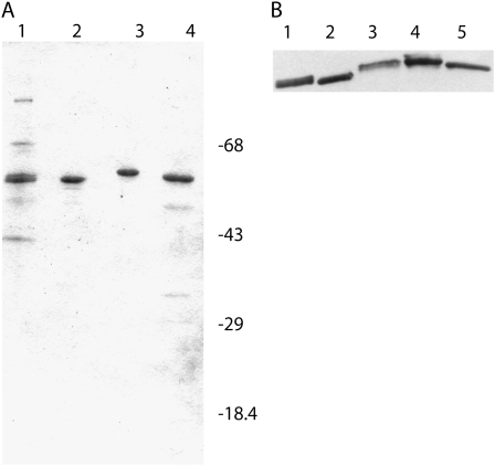
Figure 2.
SDS-PAGE and western analysis of eIF4B preparations used in in vitro translation assays. A, Each lane contains approximately 2 μg of protein. Lane 1, Native wheat eIF4B; lane 2, recombinant wheat eIF4B; lane 3, recombinant Arabidopsis eIF4B2; lane 4, recombinant Arabidopsis eIF4B1. Molecular weight markers are as indicated. The gel was stained with Coomassie Brilliant Blue. B, The polyvinylidene fluoride blot was incubated with a 1/1,000 dilution of affinity-purified Arabidopsis eIF4B2 rabbit antibodies overnight at 4°C. A 1/25,000 dilution of goat-anti-rabbit horseradish peroxidase second antibody (Kirkegaard and Perry Laboratories) was incubated for 2 h at room temperature (Browning et al., 1990). The antibody reactive bands were visualized by chemiluminescence (SuperSignal; Pierce) and exposed to film. Lane 1, Native wheat eIF4B (0.9 μg); lane 2, recombinant wheat eIF4B (0.3 μg); lane 3, native Arabidopsis eIF4B (5 μg); lane 4, recombinant Arabidopsis eIF4B2 (0.3 μg); lane 5, recombinant Arabidopsis eIF4B1 (0.3 μg).

