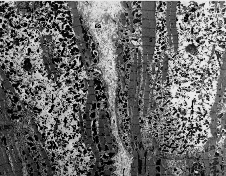
Figure 1).
Low magnification electron microscopic picture of myocardium showing typical changes in the cyosol: depleted number of contractile sarcomeres (S) and the presence of small mitochondria (arrows). The central part of the cell is occupied by glycogen (G). ES Extracellular space; N Nucleus. (Original magnification ×2600)

