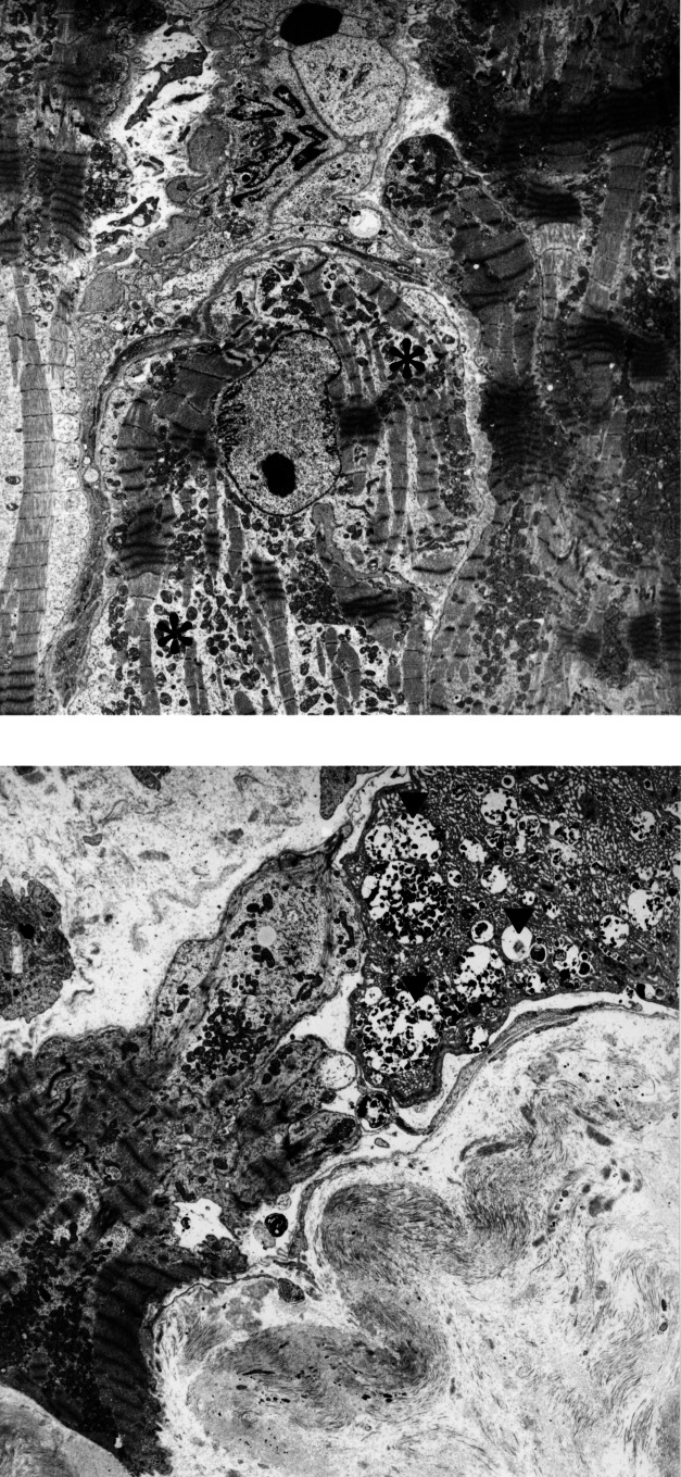
Figure 1).
Degeneration of myocytes in the failing heart. (A) Slight degeneration with partial loss of myofilaments (asterisk). (B) Severe degeneration characterized by loss of myofilaments as seen in the central myocyte. At the right upper corner, a myocyte with autophagic vacuoles (arrowheads) and areas of denatured protein are evident. This cell will most probably die an autophagic cell death. (Original magnification ×3000)

