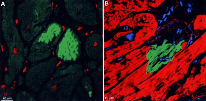
Figure 4).

Cell death by different mechanisms. (A) Storage of ubiquitin-protein complexes in two myocytes lacking a nucleus (specific fluorescence green, nuclei red). (B) Complement 9 localization indicating acute ischemic cell death (specific fluorescence green, actin staining red, nuclei and lipofuscin granules blue). Note that only one-half of the myocyte is filled with complement 9; the other part is delineated by a fine red line (remaining actin-positive structures) and contains only unspecified cytoplasm
