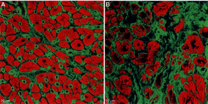
Figure 5).

The extracellular matrix stained for fibronectin (specific fluorescence green, actin staining red). (A) In normal myocardium, the amount of fibronectin is moderate and capillaries are distinctly labelled. (B) In failing myocardium, the amount of fibronectin is greatly increased and the myocytes are of different size and shape and are isolated in the fibrotic tissue
