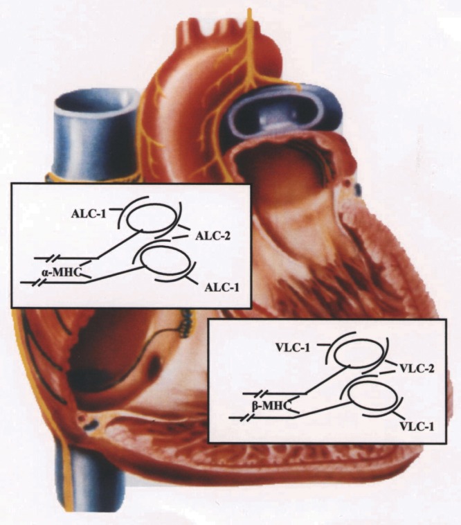
Figure 1).

Scheme of myosin heavy chain (MHC) and myosin light chain (MLC) distribution in normal human heart. On the atrial level there is α-MHC and the artrial MLC isoforms ALC-1 and ALC-2. On the ventricular level only β-MHC and the ventricular MLC isoforms VLC-1 and VLC-2 can be detected. Each myosin molecule consists of two MHCs, and on the head of each MHC there is one molecule of MLC-1 and MLC-2
