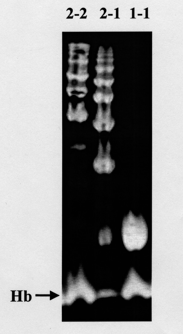
Figure 2).

Representative patterns of the different haptoglobin (Hp) phenotypes following polyacrylamide gel electrophoresis and benzidine staining of hemoglobin (Hb)-enriched serum. The upper bands correspond to Hp-Hb complexes. A band at the bottom of each lane corresponds to free Hb, and is indicated with an arrow. Hp 2-2 polymers form a series of slowly migrating bands. Hp 1-1 homodimers appear as a single rapidly migrating band. Hp 2-1 displays a mixture of slowly migrating bands and a weak band that migrates similar to the Hp 1-1 band
